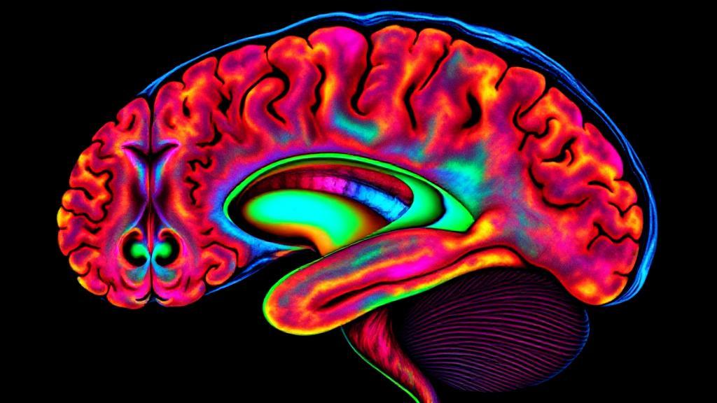
How Does a Brain MRI Work?
A brain MRI uses a strong magnetic field and radio waves to generate detailed images of the brain and brain stem. Unlike X-rays and CT scans, MRI does not use ionizing radiation, making it a safer option for repeated imaging.
The Process
-
Preparation: Patients remove metal objects and change into a hospital gown. They receive earplugs or headphones to block the machine's loud noises.
-
Scanning: The patient lies on a movable table that slides into the MRI machine. The machine's magnet aligns the protons in the body, and radio waves knock these protons out of alignment.
-
Image Creation: When radio waves are turned off, realigning protons send signals that create detailed brain images viewable from different angles.
Types of MRI Sequences
- T1-weighted images: Best for viewing anatomical structures and gray/white matter differences
- T2-weighted images: Excellent for detecting inflammation and edema
- FLAIR (Fluid-Attenuated Inversion Recovery): Particularly useful for identifying white matter lesions
- DWI (Diffusion-Weighted Imaging): Critical for early stroke detection
- MRA (Magnetic Resonance Angiography): Visualizes blood vessels and blood flow
What Can a Brain MRI Reveal?
Structural Abnormalities
- Tumors and cysts
- Brain malformations
- Hydrocephalus
- Hemorrhage
- Brain atrophy
Vascular Conditions
- Aneurysms
- Arteriovenous malformations
- Stroke
- Blood vessel blockages
Inflammatory and Degenerative Conditions
- Multiple sclerosis lesions
- Signs of neurodegenerative diseases (Alzheimer's, Parkinson's)
- Inflammation and infection
- White matter changes
Infections and Other Conditions
- Meningitis and encephalitis
- Developmental anomalies
- Traumatic brain injuries
- Abscesses and granulomas
Advantages and Limitations
Advantages
- Non-Invasive: No exposure to radiation
- Detailed Images: Superior high-resolution images
- Versatility: Can image almost any body part
Limitations
- Cost: Expensive, not always covered by insurance
- Time: Scans can take 20-60 minutes
- Claustrophobia: Anxiety due to enclosed space
- Metal restrictions: Not suitable for patients with certain metal implants
Recent Advances
Artificial Intelligence Integration
- Improve image quality
- Reduce scan times
- Enhance diagnostic accuracy
- Identify subtle patterns
Higher Field Strength
- Increased resolution
- Better contrast
- More detailed imaging
- Faster scan times
For more information, visit: